WHAT ARE FIBROIDS?
WHAT ARE FIBROIDS?
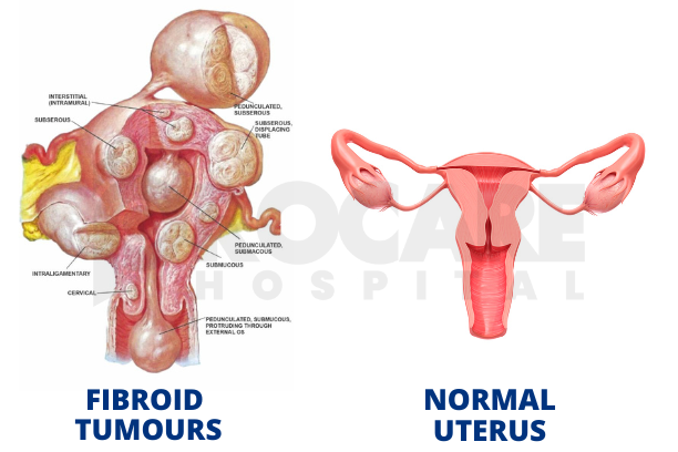
Fibroids or Uterine Fibroids are noncancerous growths or tumors of the uterus. They arise from the overgrowth of the smooth muscle (myometrium) and connective tissue of the uterus. They are also known as Uterine Myomas of Leiomyomas. Fibroids can grow on any part of the uterus. It could be inside the uterus, outside, or in the muscular layer of the uterus.
INCIDENCE
Fibroids are the most commonly diagnosed gynaecological tumour. Most common among blacks as compared with white caucasians.
RISK FACTORS
There are few known risk factors for uterine fibroids. They are:
- AGE: Fibroids develop during a woman’s reproductive years when estrogen levels are higher and tend to disappear during menopause when a woman’s estrogen levels are relatively low.
- GENETICS/HEREDITARY: Women with a family history of fibroids have a higher risk of developing fibroids.
- NULLIPAROUS WOMEN
- OBESITY
CAUSES OF FIBROIDS
The exact cause of fibroids is unknown however, research and clinical experience point to these factors:
- GENETIC PREDISPOSITION: First-degree relatives have.
- HORMONES: Two hormones produced by the ovaries. Estrogen and progesterone are increased during reproductive periods of life and stimulate the development of the uterine lining in preparation of pregnancy appears to promote fibroid growth.
CLASSIFICATION OF FIBROIDS
The growth and location of a fibroid are the main factors that determine if a fibroid leads to symptoms and problems. Different locations of the fibroids include:
- INTRAMURAL (INTERSTITIAL) FIBROIDS: This is the commonest type of fibroid. These fibroids develop within the muscular wall of the uterus. They may enlarge and distort the uterine cavity.
- SUBMUCOSAL UTERINE FIBROIDS: These fibroids develop under the lining of the uterine cavity and protrude into the cavity. These are the fibroids that have the most effect on heavy menstrual bleeding and can also cause problems with fertility and miscarriages.
- SUBSEROSAL UTERINE FIBROIDS: These fibroids originate from the serosal surface (beneath the wall) of the uterus. They can have a broad or pedunculated base and may be intraligamentary (extending between the folds of the broad ligament).
- PEDUNCULATED FIBROIDS: These are fibroids attached to the uterine wall by a stalk-like growth called outside the uterus. It is called pedunculated submucosal fibroid when inside the uterus. When outside the uterus, it is called a pedunculated subserosal fibroid.
- CERVICAL FIBROIDS: These are fibroids located in the wall of the cervix.
SYMPTOMS OF FIBROIDS
Most times, fibroids present with no symptoms. Symptoms depend on location, size and number of fibroids. Some symptoms of fibroids include:
- Heavy menstrual bleeding.
- Prolonged menstrual bleeding
- Painful menstrual period or pelvic pain.
- Abdominal swelling or mass.
- Pressure symptoms – Frequent Urination, urgency, constipation, difficult defecation, leg oedema from nerve compression.
- Backache.
- Painful Intercourse.
- Difficulty to conceive.
DIAGNOSIS OF FIBROIDS
Fibroids can be diagnosed by:
- History
- Pelvic or Abdominal Examination: The uterus will be enlarged.
- ULTRASOUND SCAN: This is the standard imaging technique to detect uterine fibroids.
- MRI: This is the most accurate imaging modality for the diagnosis of fibroids. It detects all fibroids accurately, and does precise fibroid mapping and characterization.
- HYSTEROSCOPY: This is the gold standard method to diagnose a submucous fibroid.
- LAPAROSCOPY: This is used to look for fibroids outside the uterus (subserosal fibroids) or fibroids in the layer of muscle surrounding the uterus (intramural).
TREATMENT OF FIBROIDS
Asymptomatic Fibroids may not need treatment. Symptomatic fibroid treatment options can be
MEDICAL
- NSAIDs relieve pain associated with fibroids, but this doesn’t reduce bleeding.
- TRANEXAMIC ACID: A non-hormonal medication to ease heavy menstrual period.
- GONADOTROPIN RELEASING HORMONE AGONIST (GnRHa): Helps to shrink fibroids by blocking the production of estrogen and progesterone thereby inducing a temporary menopause-like state.
SURGICAL
- MYOMECTOMY: Surgical removal of the fibroids from the uterine wall.
There are 3 types of myomectomy:
- Open Myomectomy
- Hysteroscopic Myomectomy
- Laparoscopic Myomectomy
HYSTERECTOMY: This could be partial or total removal of the uterus (womb). It remains the only proven permanent treatment for uterine fibroids. This is only considered in extreme cases.
OTHER PROCEDURES
- UTERINE ARTERY EMBOLIZATION: A non-invasive procedure that blocks blood flow to fibroids causing them to shrink.
- ENDOMETRIAL ABLATION: This is the removal of the inner lining of the uterus.
- HIGH INTENSITY FOCUSED ULTRASOUND (HIFU): ‘Is a noninvasive method. It uses ultrasound waves to generate localized heat to specifically target individual fibroids to destroy the cells.
CONCLUSION
Fibroids left untreated in severe cases can lead to complications such as infertility or problems during pregnancy such as miscarriage, pain, premature laboir and delivery, fetal growth restriction, placental abruption etc.
If you suspect you have fibroids, please speak to your gynaecologist.
Written by Dr. Stephanie, medically Verified by Dr. Fache (OBGYN).
July is Fibroid Awareness Month! Many women have uterine fibroids sometime during their lives but most women don’t know they have fibroids because they often cause no symptoms!
Throughout the month of July, @procarehospitalabuja will be running free ultrasound scans for those who want to know their fibroid status!
Knowing your status early can lead to early treatment that can increase your chance of having your dream family!
Don’t Miss this!
#procarehospital #procarehospitalabuja #fertilityclinic #hysteroscopy #ivf# #fertilityjourney #fertility #ivfjourney #gynecology #obstetrics #myomectomy #fibroids #OBGYN #urology #fertilityjourney #pediatrics #cardiology #endocrinology #internalmedicine #ivfclinic #ivfclinicinabuja #ivfinnigeria #ent #pregnancy #childbirth #breastfeeding #abujahospitals #hospitalsinabuja

